


300% up to 1500€
100% match up to €10,000
Get 225% up to 11000€
Bonus Up to €450 + 250 FS

Get: 200% up to 7760£ + 100 free spins
225% up to 7,500€
150% up to 500€ + 70 Free Spins

Get up to 7500€ +10% cashback

100% up to 450£ + 250 Free Spins
Get: 100% up to 300€
200% up to 2500€
Get 450% up to £6,000 + 325 FS
150% match up to €750/$750/£750 plus 50 free spins

Casino Welcome Bonus Up to 1500$/€/£!
150% up to 750EUR + 150 Free Spins
150% bonus up to 500€/$/£ + 50 FREE SPINS.

100% deposit match up to £500
Bonus Up to €450 + 250 FS
150% up to 3000£
400% up to 1000£ + 100 Free Spins
400% up to 2000£ + 100 Free Spins
What are the other benefits? In short, non Gamstop casinos allow UK players to enjoy a wider variety of games. This can include a broader selection of slots, table games, and even esports betting options not always found on UKGC-licensed sites. They also tend to offer more generous bonuses, faster withdrawals, and more payment options, among other perks.
What it comes down to is many players seeking the best UK online casinos without GamStop are drawn to the greater freedom these platforms offer.
Looking for a way to bypass the restrictions or explore the wider online casino scene? We’ve got you covered. Here, we’ll guide you through the safest and most reputable non Gamstop sites for 2025. We’ll also dive into everything you need to know about non Gamstop casinos; how they work, how to get the best deals, and which sites to pay attention to.
Here are some of the newest and less familiar non GamStop casinos available online for March 2025. Let’s take a look.

Betti Casino offers a competitive welcome bonus, matching 150% up to €750 on your first deposit, plus 150 free spins on Eye of Atum. A €500 deposit gets you started with €1250 to play with. The casino features a good selection of games from well-known developers like RubyPlay, Hacksaw Gaming, and Quickspin Not on Gamstop. You’ll find popular titles like Templar Tumble and Wanted Dead or Alive alongside newer releases like Potion of Madness, Go High Puppy Love, Snake Trio Bonus, and Camelot Crash.
Here’s a quick overview of what Betti offers:
Betti is a solid choice for non Gamstop gaming.
| Feature | Details |
|---|---|
| Welcome Bonus | 150% bonus up to €750 on first deposit + 150 free spins on Eye of Atum. A €500 deposit gives you €1250 to play with. |
| Game Selection | Offers a wide range of games from top developers (RubyPlay, Hacksaw Gaming, Quickspin) including Templar Tumble, Wanted Dead or Alive, and more. |
| Tournaments | Regular tournaments where you can win cash prizes and free spins. |
| Licensing | Operates as a non Gamstop casinos licensed by the Curaçao Gaming Control Board. |
| Banking | Minimum deposit €20, maximum deposit €5000; supports Visa, Mastercard, Bank Transfer, Ethereum, Bitcoin, and Litecoin. |
| Payouts | Aims to process withdrawals within two days (processing times may vary). |
| Overall Experience | A solid choice for non GamStop gaming with competitive bonuses and flexible banking. |

JettBet Casino offers a large welcome bonus spread across your first four deposits, totaling 450% up to €6000, plus 425 free spins. With over 3100 games, including slots, roulette, and blackjack, JettBet emphasizes fairness, stating that its games are officially tested, approved, and certified. Popular titles include African King Hold’N’Link, Big Catch Bonanza, Moneyfest, and Book Of Ra. The game library features titles from developers like Netgame, Popiplay, Novomatic, Mascot, and others.
Here’s a quick overview of what JettBet offers:
| Feature | Details |
|---|---|
| Welcome Bonus | 450% bonus up to €6000 across four deposits plus 425 free spins. |
| Game Selection | Over 3100 games including slots, roulette, and blackjack; titles like African King Hold’N’Link, Big Catch Bonanza, Moneyfest, and Book Of Ra. |
| Promotions | Additional promotions available, including free spin bonuses. |
| Licensing | Non Gamstop casino licensed by the Curaçao Gaming Control Board. |
| Developers | Games provided by Netgame, Popiplay, Novomatic, Mascot, and others. |
| Banking | Accepts Visa, Mastercard, e-wallets, and bank transfer (cryptocurrency not supported). |
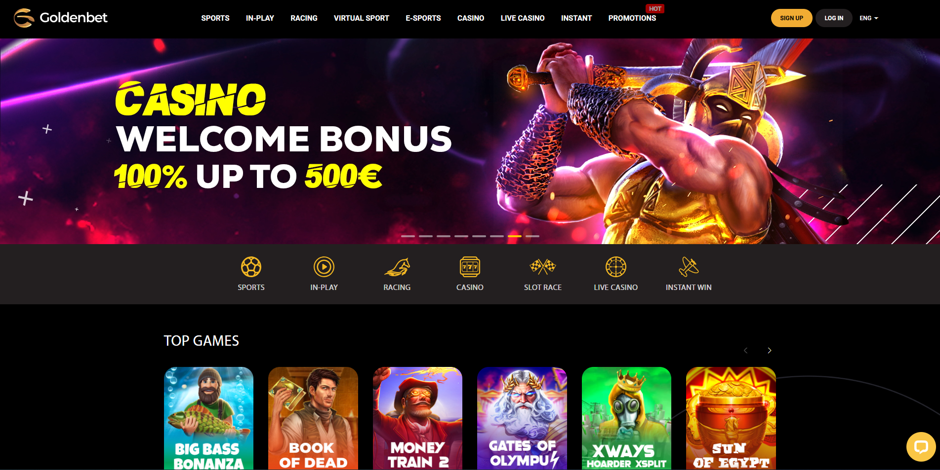
GoldenBet Casino has earned a solid 4.2/5 rating from almost 2,000 reviews on Trustpilot, which speaks to its reputation. They also offer a 100% match bonus up to €500 (or the equivalent in other currencies they accept) when you make your first deposit. Just keep in mind, you’ll need to deposit at least €20 to qualify, and the bonus is specifically for their slots. Game-wise, they’ve got popular titles like Book of the Dead, Sun of Egypt 2, Money Train 2, Legacy of Dead, and Hand of Anubis, all powered by well-known developers like Relax Gaming, Hacksaw Gaming, Play’n Go, and Quickspin.
Here’s a quick overview of what GoldenBet offers:
| Feature | Details |
|---|---|
| Rating | Solid 4.2/5 on Trustpilot from nearly 2,000 reviews |
| Welcome Bonus | 100% match bonus up to €500 on first deposit (slots only) |
| Game Selection | Popular titles like Book of the Dead, Sun of Egypt 2, Money Train 2, Legacy of Dead, Hand of Anubis |
| Developers | Games from Relax Gaming, Hacksaw Gaming, Play’n Go, Quickspin, and others |
| Licensing | Operates under a Curaçao license |
| Banking | Minimum deposit of €20; accepts Visa, Mastercard, and e-wallets |
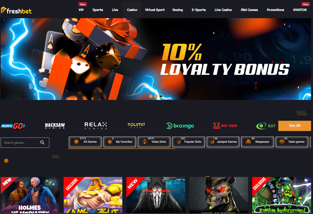
Freshbet Casino has a pretty enticing welcome offer: a 150% match bonus up to £1500. Just go to your profile when you make your first deposit (at least £20), and you’ll get the bonus right away. They’ve got a decent selection of games, including popular ones like Book of the Dead, Sweet Bonanza, Great Rhino, and Book of Ra Deluxe, plus some newer titles like Bigger Bass Bonanza and Sweet Alchemy 100. You’ll find games from some of the big names in the business, like Pragmatic Play, NetEnt, Play’n Go, and Quickspin.
Here’s a quick rundown of what Freshbet offers:
| Feature | Details |
|---|---|
| Welcome Bonus | 150% match bonus up to £1500 on first deposit (minimum deposit: £20) |
| Game Selection | Popular titles include Book of the Dead, Sweet Bonanza, Great Rhino, Book of Ra Deluxe, Bigger Bass Bonanza, and Sweet Alchemy 100 |
| Developers | Powered by Pragmatic Play, NetEnt, Play’n Go, and Quickspin |
| Licensing | Operated by Ryker B.V. and licensed in Curaçao |
| Banking | Minimum deposit of £20; accepts Visa, Mastercard, e-wallets, and cryptocurrencies |

1Red Casino offers a substantial welcome bonus, giving you a 200% match on your first deposit up to £7760 , plus 100 free spins. You’ll need to deposit at least £20 to qualify for this offer. Their game selection includes popular titles like Thunder Coins, Fishin’ Frenzy The Big Splash, 3 Volcano Coins, Book of Ra, and BlackWolf 2, alongside newer additions such as Happy Fish, Lightning Clovers, and Sheriff Chase. BassWin also features a live casino with games like live roulette and Golden Land.
Here’s a quick overview of what 1Red offers:
| Feature | Details |
|---|---|
| Welcome Bonus | 200% match bonus up to £7760 plus 100 free spins on first deposit |
| Game Selection | Popular slots including Thunder Coins, Fishin’ Frenzy The Big Splash, 3 Volcano Coins, Book of Ra, BlackWolf 2, plus newer titles like Happy Fish, Lightning Clovers, Sheriff Chase; also features a live casino with roulette and Golden Land |
| Licensing | Registered and established under the laws of Curaçao |
| Banking | Minimum deposit of £20; accepts Visa, Mastercard, Apple Pay, and cryptocurrencies |

Donbet Casino offers a welcome package of up to £750 plus 50 free spins, with a 40x wagering requirement on the bonus. Their game library features popular titles like Big Big Fishing Fortune, Clovers of Fortune, Crazy Tikis, Big Bass Bonanza, and Gonzo’s Quest. If you have any questions or run into any problems, their support team is available 24/7.
Here’s a quick rundown of what DonBet offers:
| Feature | Details |
|---|---|
| Welcome Package | Up to £750 plus 50 free spins with a 40x wagering requirement |
| Game Library | Includes Big Big Fishing Fortune, Clovers of Fortune, Crazy Tikis, Big Bass Bonanza, and Gonzo’s Quest |
| Support | 24/7 customer support available |
| Licensing | Licensed and regulated in Curacao |
| Banking | Accepts Visa, Mastercard, Apple Pay, PayPal, and various e-wallets (crypto not accepted) |
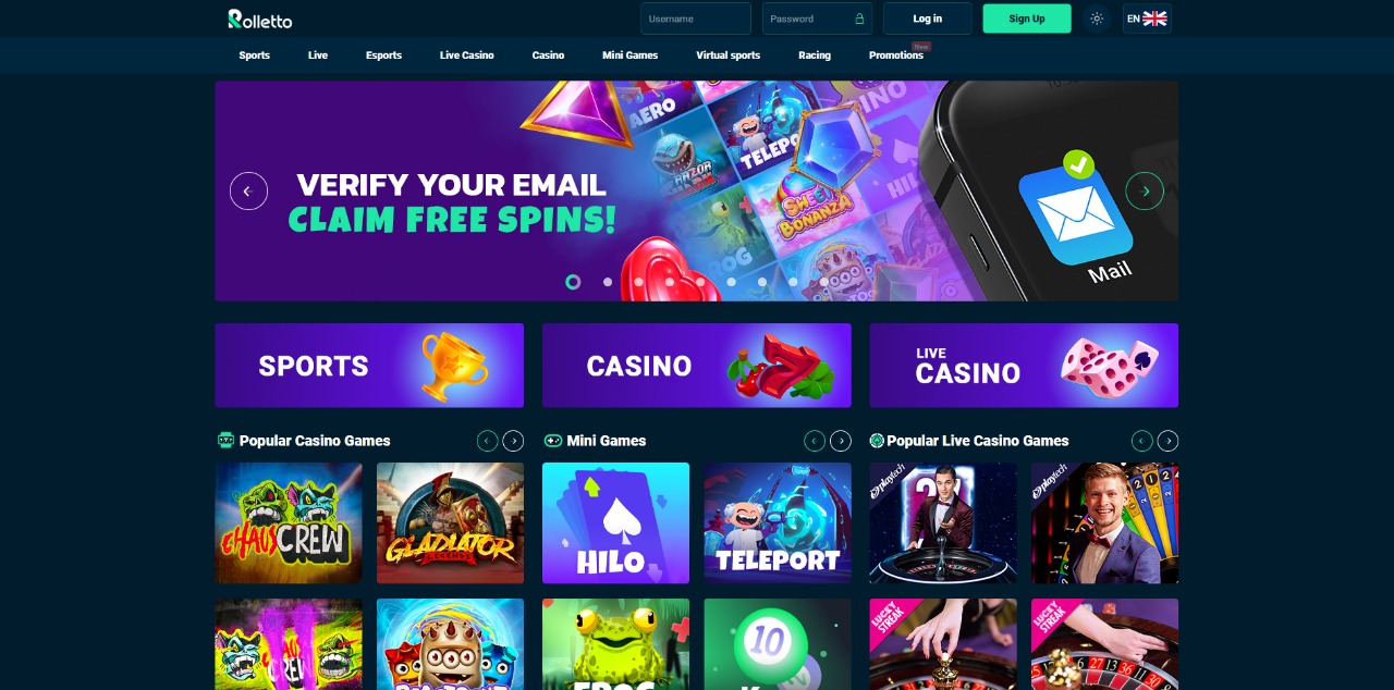
Rolletto Casino offers an attractive, tiered welcome bonus. A 50% match bonus up to £500 plus 50 free spins is available for deposits between £20 and £500 (or equivalent currency). For high rollers, deposits between £500 and £1000 (or equivalent currency) will receive a 100% match bonus. The game selection at Rolletto Win includes popular titles such as Aztec Magic Megaways, their exclusive Rolletto King, Magic Piggy, and Holmes vs Moriarty, provided by reputable developers like BGaming, Hacksaw Gaming, Slotopia, Quickspin, and Playson.
Here’s a quick overview of what Rolletto provides:
| Feature | Details |
|---|---|
| Bonus | 50% match bonus up to £500 + 50 free spins for deposits of £20-£500, or 100% match bonus for deposits of £500-£1000 |
| Licensing | Licensed in Curaçao |
| Banking | Accepts Visa, Mastercard, Apple Pay, PayPal, and several popular cryptocurrencies |
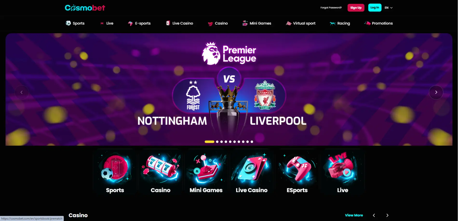
Cosmobet Casino welcomes you with a cosmic welcome bonus. Deposits between £20 and £500 (or equivalent currency) receive a 150% bonus plus 50 free spins. High-rollers depositing between £500 and £1000 (or equivalent currency) get a 100% bonus. Cosmobet features popular games like Aztec Magic Megaways, Evil Eyes Dragon Shrine, and Holmes vs Moriarty, plus exclusive titles like Cosmobet Diamonds Hunt and The Cosmobet Greatest Catch. Games are from developers like BGaming, Hacksaw, Slotopia, and Exoplay, among others.
Here’s a quick overview:
| Feature | Details |
|---|---|
| Bonus | 150% bonus up to £500 + 50 free spins for deposits of £20-£500, or 100% match bonus for deposits of £500-£1000. |
| Game Selection | Offers popular games like Aztec Magic Megaways, Evil Eyes Dragon Shrine, and Holmes vs Moriarty, plus exclusive titles like Cosmobet Diamonds Hunt and The Cosmobet Greatest Catch. |
| Licensing | Licensed in Curaçao. |
| Banking | Accepts Visa, Mastercard, Apple Pay, PayPal, and various cryptocurrencies. |

MyStake Casino, a casino not on GamStop, welcomes new players with a 300% Welcome Package worth up to £1,500 in bonus money for casino slot games. This package is spread across your first three deposits: a 150% bonus up to £750 on your first deposit, a 100% bonus up to £500 on your second, and a 50% bonus up to £250 on your third. MyStake features popular games like Book of Dead and Gates of Olympus, plus exclusive titles like Gates of Mystake, CandyRush, and MyStake Greatest Catch. Games are provided by proven developers like BGaming, Hacksaw, Slotopia, and Exoplay.
Here’s a quick overview:
| Feature | Details |
|---|---|
| Welcome Bonus | 300% Welcome Package up to £1,500 across your first three deposits (150% up to £750, 100% up to £500, 50% up to £250) |
| Game Selection | Popular slots like Book of Dead and Gates of Olympus, plus exclusive titles such as Gates of Mystake, CandyRush, and MyStake Greatest Catch |
| Developers | Games provided by BGaming, Hacksaw, Slotopia, and Exoplay |
| Licensing | Licensed in Curaçao |
| Banking | Accepts Visa, Mastercard, and various cryptocurrencies |

VeloBet offers a two-tiered welcome bonus. Deposits between £10 and £500 (or equivalent currency) will receive a 150% match bonus plus 70 free spins. For larger deposits between £500 and £1000 (or equivalent currency), the bonus is a 100% match plus 70 free spins on Play’n GO’s Book of Dead. Use promo code WELCOME when depositing to claim your free spins, which are valid for 24 hours. VeloBet features popular games like Magic Piggy, Aztec Magic Megaways, Sun of Egypt 2, Solar Queen, and Wanted Dead or a Wild, along with exclusive titles such as Velobet Diamonds Hunt and Candy Splash Velobet.
Here’s a quick overview:
| Feature | Details |
|---|---|
| Bonus | Two-tiered welcome bonus: 150% + 70 free spins for deposits £10-£500; 100% + 70 free spins for deposits £500-£1000 (on Book of Dead, use promo code WELCOME; free spins valid for 24 hours) |
| Game Selection | Offers popular games like Magic Piggy, Aztec Magic Megaways, Sun of Egypt 2, Solar Queen, and Wanted Dead or a Wild; exclusive titles include Velobet Diamonds Hunt and Candy Splash Velobet |
| Licensing | Non GamStop casino under a Curaçao license |
| Banking | Accepts Visa, Mastercard, e-wallets, and various cryptocurrencies; minimum withdrawal is 20€ (or equivalent), with maximum withdrawal limits varying by payment method |
| Casino | Best For | Promotion | Minimum Deposit |
|---|---|---|---|
| Betti Casino | Slots | 150% bonus up to €750 on first deposit + 150 free spins on Eye of Atum | €20 |
| JettBet Casino | Slots, Roulette & Blackjack | 450% bonus up to €6000 across four deposits + 425 free spins | Not specified |
| GoldenBet Casino | Slots | 100% match bonus up to €500 on first deposit (slots only) | €20 |
| Freshbet Casino | Slots | 150% match bonus up to £1500 | £20 |
| 1Red Casino | Slots, Table Games & Live Casino | 200% match bonus up to £7760 plus 100 free spins on first deposit | £20 |
| Donbet Casino | Slots | Welcome package up to £750 plus 50 free spins (40x wagering requirement) | Not specified |
| Rolletto Casino | Slots | 50% match up to £500 + 50 free spins (deposits £20-£500) or 100% match bonus (deposits £500-£1000) | £20 |
| Cosmobet Casino | Slots | 150% bonus up to £500 + 50 free spins (deposits £20-£500) or 100% bonus (deposits £500-£1000) | £20 |
| MyStake Casino | Slots | 300% Welcome Package up to £1,500 across three deposits (150% up to £750, 100% up to £500, 50% up to £250) | Not specified |
| VeloBet | Slots | 150% bonus + 70 free spins (deposits £10-£500) or 100% bonus + 70 free spins on Book of Dead (deposits £500-£1000). Use promo code WELCOME. | £10 |
Gamstop is a self-exclusion program designed to help UK players control their gambling habits. Operated by the UK Gambling Commission (UKGC), it allows individuals to voluntarily block themselves from all UK-licensed casino and betting sites for 6 months, 1 year, or 5 years.
Once registered, players cannot access any UKGC-regulated gambling sites until the exclusion period ends. GamStop restrictions apply to all licensed platforms, preventing users from creating new accounts, depositing funds, or placing bets. However, since the system only covers UK-based operators, it does not affect offshore casinos, allowing players to gamble at casinos not on GamStop without restrictions.
Players choose non Gamstop online casinos for many reasons. One major reason is to regain access to online gambling after self-exclusion. Introduced in 2018 by the UK Gambling Commission (UKGC), GamStop was designed to help problem gamblers by blocking access to all UK-licensed betting sites for a set period—ranging from six months to five years. The program has helped many people, however, it’s not perfect. Namely, many players find its blanket restrictions too rigid, particularly if they believe they have regained control over their gambling habits.
Since GamStop cannot be reversed once activated, even players who feel ready to return must wait until their exclusion period ends. This can be frustrating, and casinos not on Gamstop provide an alternative, allowing you to resume play.
However, re-entry to online gambling isn’t the only reason to opt for casinos Not on Gamstop. Another major draw is that these sites are non-UK casinos, which means more variety – different games, bonuses, currencies, and so on. Not all players want to use the same old sites all the time, and in that way, non Gamstop casinos are a good change of pace.
Here are the main reasons players like non GamStop casinos:

We look closely at every casino site Not on Gamstop we recommend, using a selection criteria we’ve honed over the years. The main areas we’re interested in are licensing, security, and user reviews.
Let’s start with licensing. Just because a site isn’t registered with the UK Gambling Commission doesn’t mean it’s a lawless, wild-west style site. Non Gamstop casinos still adhere to strict industry standards, they just fall under different licensing than UK casinos. Typically non Gamstop casinos are licensed with the Malta Gaming Authority (MGA) or the Curaçao Gaming Control Board. We ensure that every site on our list is registered under a reputable licensing body.
Security is essential for any website where you’ll be sharing your details and spending your money. We check the site uses appropriate industry standard encryption and has safe data handling practices (for example, using gateways for payments, and not taking any more details than strictly necessary).
User reviews are also a crucial part of our evaluation process. We don’t just take a casino’s word for it – we want to know what real players are saying. We look at user reviews for consistent themes and patterns in feedback. We’re interested in everything from the fairness of the games and the speed of payouts to the helpfulness of customer support and the overall user experience. Are players happy with the game selection? Do they find the site easy to navigate? Have they encountered any issues with withdrawals or account verification? By carefully analyzing user reviews, we can get a genuine sense of what it’s like to play at each non GamStop casino, allowing us to identify any potential red flags and ensure we’re recommending sites that prioritize player satisfaction.
User reviews are also one of the reasons our recommendations can change from year to year. Some casinos may start strong but then experience issues they fail to correct in a timely way. At the same time, new casinos break out into the non GamStop market and if they follow our criteria, we think they deserve to be highlighted too.
Non GamStop casinos and GamStop casinos have a few differences, and these differences can help you determine which one is right for you.
Let’s start with the biggest difference. As the name suggests, non Gamstop casinos are not part of the GamStop exclusion scheme. GamStop is a free tool that allows people in the UK to exclude themselves from all licensed gambling websites and apps for a set period of time (six months to five years). Crucially, all online casinos licensed by the UKGC must be part of the GamStop scheme.
So essentially, if you’ve signed up for GamStop then all UK casinos are now out of reach for you until the exclusion period ends, with no exceptions. And while the testimonials on the GamStop website attest to how helpful the scheme can be, that doesn’t mean it’s without its flaws. For example, the minimum time you can self-exclude is six months, which is overly restrictive for many players. Some people just want to be excluded for a few weeks or a month but find themselves locked out of gambling sites for far longer, even if they’re not displaying any risky behavior. Non Gamstop casinos put you back in the driver’s seat – if you feel ready to play again, you can!
However, it’s not all about GamStop. Non Gamstop casinos have other benefits too. Since they’re operating outside the UKGC’s jurisdiction, they often come with fewer restrictions on gameplay, bonuses, and payment methods. UKGC-licensed casinos have strict limits on deposit amounts, wagering requirements, and even features like autoplay and spin timers for slots. Casinos not on Gamstop don’t have to play by these rules. This means higher deposit limits, bigger bonuses, and faster gameplay, giving you more freedom over how you bet.
Another major difference is payment flexibility. UKGC casinos have banned credit card deposits altogether, while non Gamstop casinos still allow them. Most also accept cryptocurrencies like Bitcoin, Ethereum, and Litecoin. Cryptocurrency is a dealbreaker for some players, especially if you value speed, low fees, and privacy. Crypto transactions are faster than bank transfers, often processing within minutes. They also offer more anonymity, and many non GamStop casinos reward crypto users with exclusive bonuses.
Then there’s the game selection. Non GamStop casinos often have a broader range of games because they’re not restricted to UK-approved providers. This means access to a wider variety of slots, live dealer games, and table games from top international developers. Some also offer sports betting, virtual sports, and eSports betting—markets that can be more limited at UKGC-regulated sites.
Of course, there are trade-offs. UKGC casinos come with stronger consumer protections, stricter responsible gambling measures, and access to UK-based dispute resolution services. Non GamStop casinos may not offer the same level of regulatory oversight when it comes to complaint handling or enforcing responsible gambling tools.
Ultimately, the choice comes down to what you value most. If you want a tightly regulated environment with built-in safeguards, UKGC casinos are the way to go. But if you prefer more control over your gambling experience, better bonuses, and fewer restrictions, non Gamstop casinos come out on top.
| Feature/Aspect | Non Gamstop Casinos | GamStop Casinos |
|---|---|---|
| Exclusion Scheme | Not part of GamStop; flexible, allowing you to resume play at any time | Mandatory self-exclusion via GamStop (minimum six months to five years) |
| Gameplay & Bonuses | Fewer restrictions, higher deposit limits, bigger bonuses, and faster gameplay | Strict limits on deposits, wagering requirements, and gameplay features |
| Payment Methods | Accept credit cards and cryptocurrencies for faster, more flexible transactions | Credit card deposits banned; fewer payment options available |
| Game Selection | Broader variety including slots, live dealer games, table games, sports betting, and eSports | Limited to UK-approved providers with a narrower game range |
| Regulatory Oversight | Operate outside UKGC jurisdiction; fewer consumer protections and responsible gambling measures | Strong UKGC oversight with enhanced consumer protection and dispute resolution services |
For UK players seeking more freedom and fewer restrictions, casinos not on GamStop provide a flexible alternative to traditional UK-licensed platforms. These gambling sites are typically offshore casinos, licensed outside the UK, and are not part of the GamStop self-exclusion scheme—making them accessible to self-excluded users.
Whether you’re looking for non-UK casinos, sites with fast withdrawal options, or platforms that support PayPal deposits, there’s a wide variety of non-GamStop casinos to choose from. Each type offers different features, from international game libraries and crypto payments to larger bonuses and streamlined registration.
In this guide, we’ll break down the most popular types of non GamStop casinos and help you decide which is right for your gaming style and preferences.
These are international online casinos operating outside the United Kingdom’s jurisdiction. Non UK Casinos hold licenses from offshore authorities, allowing them to accept UK players without adhering to UK Gambling Commission regulations. This independence enables them to offer unique games, promotions, and payment methods not typically found in UK-regulated casinos.
Pros:
Cons:
Fast Withdrawal Non Gamstop casinos prioritize quick processing times for withdrawals, ensuring players receive their winnings promptly. By streamlining verification processes and utilizing efficient payment methods, they enhance the overall gaming experience.
Pros:
Cons:
Offshore casinos are online gambling platforms based in jurisdictions outside the UK. They operate under licenses from international regulatory bodies and cater to a global audience, including UK players seeking alternatives to UKGC-regulated sites.
Pros:
Cons:
Paypal casinos Not on Gamstop offer PayPal as a payment method, providing a familiar and secure option for UK players. PayPal’s reputation for safety and convenience makes these casinos appealing to those seeking reliable deposit and withdrawal options.
Pros:
Cons:
One of the biggest draws of casinos not on GamStop is the sheer variety of games, and slots take center stage. You’ll find everything from classic fruit machines with simple gameplay to the latest video slots packed with bonus features and fun graphics. Think popular titles like Big Bass Bonanza – everyone loves a good fishing-themed slot – or Book of Dead, with its Egyptian theme and the potential for some serious wins. Money Train 2 is another common find, and Gates of Olympus is a great choice if you enjoy Greek mythology-themed games. Honestly, the list goes on and on.
And it’s not just slots. You’ll usually find all the classic table games like blackjack, roulette, and baccarat, often with a few different versions to keep things interesting.
A lot of what makes a good game comes down to the developers. Casinos not on GameStop often work with some of the biggest names in the business, like NetEnt, Pragmatic Play, and Play’n GO – they’re known for making consistently good games that prioritize fairness and security. Relax Gaming is another developer that commonly works with casinos not on Gamstop; they’re the brains behind the Money Train series and a bunch of other innovative titles. But it’s not just the big guys. You’ll also see games from smaller studios that are coming up with some creative stuff.
UK players who register at casinos not on GamStop gain access to some of the most exciting and high-paying online slots—many of which aren’t available on UK-licensed platforms. These non-GamStop slots often come with fewer restrictions, larger win potential, and exclusive features. Below are some of the most popular and trusted slot games that you can enjoy at non-GamStop casinos, including some we’ve fully reviewed on LGC.
Starburst Not on Gamstop remains a fan favourite due to its clean graphics, cosmic theme, and fast-paced gameplay. This low-volatility slot pays both ways and features expanding wilds that trigger re-spins, offering consistent action without draining your balance. Perfect for new players or casual spins at PayPal-friendly non-GamStop casinos.
Rich Wilde’s ancient Egyptian adventure is a go-to for high-risk, high-reward slot fans. Book of Dead Not on Gamstop offers a thrilling free spins round with a special expanding symbol that can lead to full-screen wins. It’s high volatility, big win potential, and cult following make it a top pick among UK players outside GamStop.
With its “Pay Anywhere” mechanic, cascading reels, and massive multipliers (up to 500x), Gates of Olympus Not on Gamstop is one of the most explosive slots not on GamStop. Zeus can drop random multipliers after any spin, and the free spins round can deliver huge wins—making it a must-try for players chasing adrenaline.
An iconic slot that introduced the Avalanche™ mechanic, Gonzo’s Quest takes you on a hunt for El Dorado. With every consecutive win, the multiplier increases—up to 5x in the base game and 15x during free falls. Its immersive visuals and smooth gameplay make it a timeless choice for UK players on non-GamStop platforms.
Fishing meets fun in Big Bass Bonanza, a popular non-GamStop slot with light-hearted graphics and big win potential. Land 3 or more scatters to trigger free spins where fish symbols hold cash values and the Fisherman wild collects them. With retriggers and multiplying wilds, this slot keeps things reely exciting.
This is by no means an exhaustive list, so you’ll come across plenty of games from other developers too. Most non GamStop casinos list the developer when you hover over or click through to the game (it’s often under the title). At Casinos not on Gamstop you will find also popular software like Quickspin, Playn’Go, Netent and many more.
Non Gamstop casinos are known for their competitive bonuses, designed to attract and reward players. Welcome bonuses are a common feature, and these can be quite generous. For example, Betti Casino offers a 150% match up to £750 plus 150 free spins, giving you a substantial boost to your initial bankroll. JettBet goes even further, spreading a 450% bonus up to £6000 plus 425 free spins across your first four deposits. Donbet offers a 150% match up to £500 and 200 free spins, while Freshbet offers a massive 150% match.
Tiered bonuses are another popular approach. Rolletto offers a 50% match up to £500 for smaller deposits, and a 100% match for larger ones. Cosmobet uses a similar strategy, offering 150% or 100% matches depending on the deposit amount. VeloBet also offers a tiered welcome bonus, with extra free spins thrown in.
Free spins are often bundled with deposit bonuses, as seen at Betti, JettBet, Freshbet, 1Red, Rolletto, Cosmobet, and VeloBet. These give you a chance to try out slots risk-free. While not all casinos not on Gamstop advertise cashback or loyalty programs as prominently as the welcome offers, they are sometimes available. It’s always a good idea to check the promotions page of the casino you’re interested in to see the full range of offers. And remember, all bonuses come with terms and conditions, including wagering requirements, which you should always review before claiming any offer.
Bonus terms and conditions vary slightly from casino to casino, but for the most part, they are almost identical. That’s why we’ve compiled a helpful list below of what you can generally expect. Please note that these aren’t all the T&Cs, just the key points to be aware of:
So, we’ve covered some top new casinos not on Gamstop, but that doesn’t mean your options end there. The world of online casinos not on GamStop is constantly evolving, with new sites popping up regularly. Here are a few more worth checking out:
This is just a small taste of what’s out there – the non Gamstop casino scene is thriving, so if you’re looking for even more options, there are plenty to discover.
NonGamstop casinos offer a variety of payment options to suit different players. You can typically use major debit cards like Visa and Mastercard, which are widely accepted for both deposits and withdrawals.
For faster transactions, many sites support e-wallets like MiFINITY and AstroPay, which provide quick and secure payments without sharing your banking details.
If you prefer direct bank transfers, most non Gamstop casinos allow bank wire deposits and withdrawals, though these can take longer to process. Lastly, most (in our experience, all) non Gamstop casinos also accept cryptocurrencies like Bitcoin, Ethereum, Tether, and Litecoin. Which specific cryptocurrencies they will accept can vary, but Bitcoin and Ethereum are usually a given.
casinos not on Gamstop offer plenty of benefits but also have some drawbacks. Let’s look at the pros and cons of sites not on GamStop.
These are the main reasons to opt for non Gamstop gambling:
These are the main reasons non Gamstop gambling might not be for you right now:
Casinos not on GamStop understand that many players prefer the convenience of mobile gaming. Therefore, they typically offer ways to play on your smartphone or tablet. Some sites may have dedicated mobile apps you can download and install on your device. These apps are often designed to provide a smoother and more tailored gaming experience.
More commonly, these casinos will have websites optimized for mobile browsers. This means the website automatically adjusts its layout and design to fit your smaller screen, making it easy to navigate and play games without needing to download anything.
Yes. Typically non GamStop casinos are licensed with the Malta Gaming Authority (MGA) or the Curaçao Gaming Control Board (CGCB), although occasionally you will see a casino licensed elsewhere. Crucially, the MGA and CGCB license boards are reputable and widely respected outside the UK.
For example, Malta is widely considered the home of European iGaming, and the MGA is one of the most well-respected bodies in the global gambling industry. It is committed to fair and lawful gambling practices. To drive this home, GamStop gambling sites you’ll recognize in the UK are usually also under MGA outside of UK borders – for example, WilliamHill is registered under the Malta Gaming Authority.
The main difference between the UKGC and the MGA is simply how they grant licenses – broadly the UK has two types of licenses, while the MGA has four. These differences aren’t really important from a player’s perspective, since they mostly pertain to things like the specific types of gambling offered (e.g., software provision vs. specific game categories like lotteries or skill-based betting) and the operational aspects of running a gambling business, rather than the player’s experience of fairness or security.
The Curaçao Gaming Control Board (CGCB) is another significant licensing body you’ll often see. It’s a long-standing regulator, established in 1996, overseeing gambling operations within Curaçao. While it may not have the same prestige as the MGA, it still holds a significant presence. From a player’s perspective, a Curaçao license indicates that the casino has met certain requirements for financial stability and responsible business practices, which are important for ensuring your funds are safe. While some players might perceive Curaçao licenses as less stringent, they still provide a level of oversight.
Beyond these two, you might occasionally encounter casinos licensed in other jurisdictions like Costa Rica or Panama. However, these are less common and generally viewed with more caution by players, as these jurisdictions often have less stringent regulatory oversight compared to Malta or Curaçao. It’s always wise to research any licensing body you’re unfamiliar with before playing at a casino.
In short, while these boards are slightly less stringent than the UKGC, they mostly just approach licensing differently.
In short, yes. Most reputable casinos not on Gamstop understand the importance of responsible gambling and offer various tools to help players stay in control. A common feature is the ability to self-exclude by contacting customer support. This isn’t like the nationwide GamStop system, but it does allow you to take a break from that particular casino. Many sites also let you set deposit limits so you can manage your spending. Time-out features are also common, letting you temporarily suspend your account if you feel that’s necessary.
Besides these tools, many non UKGC casinos offer information and resources on responsible gambling. They might have dedicated pages explaining their policies, giving tips for safe gambling, and linking to international support organizations. If you’re in the UK and feel you would benefit from gambling help, you can get free help from GambleAware and GamCare. The UK Gambling Commission also keeps an updated list of organizations that can help.
Here’s the bottom line. Casinos Not on Gamstop are regulated differently than UK casinos, but this doesn’t mean they don’t care about responsible gambling. Most realize that player well-being is important, and they try to create a safe environment where you can enjoy yourself and want to return. While it’s ultimately up to each player to be responsible, the fact that these casinos offer self-exclusion, deposit limits, and access to support shows they take responsible gambling seriously.
If you’re a UK player looking for online gambling options, non-Gam-Stop casinos offer a different experience. GamStop is a valuable tool for those struggling with problem gambling, but its blanket restrictions can sometimes feel too much. And for others, they’re just unnecessary. Non-Gam-Stop sites provide an alternative, and for many players, they offer some real advantages. Think more flexibility in your gameplay, a wider selection of games, and often, features like accepting cryptocurrency.
If you want to try a casino not on Gamstop, start with the ones in this list. We vet the casinos we recommend to ensure they are under a reputable licensing body and have good customer support and security practices. Go have fun and remember to gamble responsibly!
Here we’ll answer some rapid-fire questions about non Gamstop casinos. You can use this section as a quick guide to everything you need to know about these platforms.
A: Yes. As long as you choose a reputable non Gamstop casino that’s licensed in a trusted jurisdiction, like Curacao or Malta, you’re safe. Look for casinos with strong security measures, like encryption and fair game practices, to ensure a secure experience.
A: Yes. While non Gamstop casinos aren’t part of the UK’s official self-exclusion scheme, many of them offer their own responsible gambling tools. You can still choose to self-exclude from these sites directly through their customer support or account settings.
A: Yes, using these casinos doesn’t stop you from using any other casinos. However, if you’ve opted into GamStop, you won’t be able to access UKGC-licensed sites until your exclusion period ends.
A: Yes. Many non Gamstop casinos accept the same payment methods as UKGC-licensed casinos, including credit/debit cards, e-wallets, and even cryptocurrencies. Check the casino’s payment options to see what works best for you.
A: Yes, in many cases, non GamStop casinos offer a broader range of games, including international slots, table games, and live dealers that may not be available on UKGC-licensed platforms.
A: Yes, non Gamstop casinos are legal to use in the UK, they’re just not regulated by the UK Gambling Commission (UKGC). However, it’s important to note that some sites put restrictions on users from certain countries. For example, they may ban users from the US, UK, or other countries where gambling laws are stricter. Still, it doesn’t make it illegal to use those sites and some users still choose to use them with a VPN.
A: Look for licensing information, read user reviews, and check the casino’s security features. A trustworthy non Gamstop casino will have proper licensing, secure payment options, clear terms for bonuses, and a reputation for paying out winnings on time.
A: Yes, most non Gamstop casinos offer customer support through live chat, email, or phone. Make sure to check the availability and responsiveness of their support team before signing up to ensure you can get help when needed.